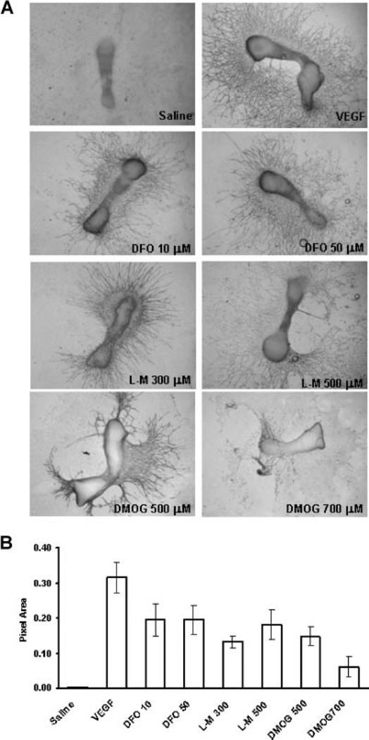
Figure 2.
PHD inhibitors induce functional angiogenesis. (A) CD31 staining. Metatarsals were dissected from 17.5-day mouse embryos. They were cultured in MEM. PHD inhibitors at the concentrations indicated, saline, or VEGF (10 ng/mL) were applied for 24 h. Subsequent capillary sprouting is assessed by CD31 staining. Robust sprouting is observed for the treatment and positive control groups. (B) Image analysis. Pixel area of capillary sprouts identified by CD31 staining was measured using Image J. The graph represents combined results from three separate experiments.

