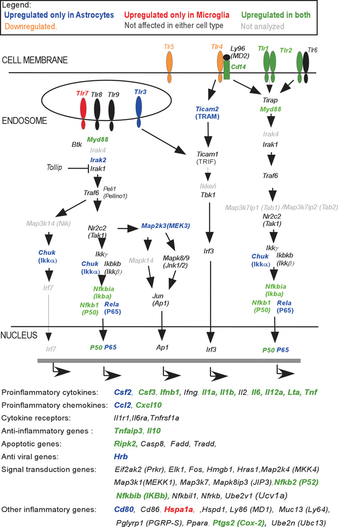
Fig. 2. Changes in mRNA expression following TLR7 and/or TLR9 agonist stimulation in astrocytes and microglia.
Cultured astrocytes and microglia were stimulated with 5 µM imiquimod or 80 nM CpG-ODN 1826 or both. RNA was isolated at 6 hps, reverse transcribed and cDNA was analyzed for mRNA expression by quantitative real-time PCR analysis using TLR signaling pathway array. Genes are presented as a schematic of their protein’s involvement in the TLR signaling cascade or as genes transcribed following the signaling cascade. Increased mRNA expression of these genes in both microglia and astrocytes is indicated by green lettering, in astrocytes only by bold blue lettering and in microglia only by bold red lettering. Tlr4 mRNA was downregulated in microglia, while Tlr5 mRNA was downregulated in both microglia and astrocytes. Genes indicated in black lettering were not altered in either cell type and in grey lettering were not analyzed for mRNA expression.

