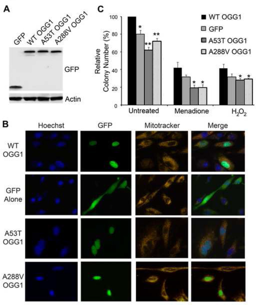
Fig. 6. Cells expressing the OGG1 variants are sensitive to DNA damaging agents and have decrease colony formation.
(A) OGG1−/− MEFs were transfected with the indicated GFP-fusion proteins and 24 hrs later cells were lysed, run on a 10% polyacrylamide gel and immunoblotted with anti-GFP antibodies to show expression of the constructs. Actin was used as a protein loading control. (B) OGG1−/− MEFs were transfected with the indicated GFP-fusion proteins. 24 hrs later, cells were incubated with Mitotracker and Hoechst stains and imaged live to determine the cellular localization of the GFP-tagged WT and polymorphic OGG1 proteins. (C) OGG1−/− MEFs were transfected with GFP-tagged constructs containing WT or OGG1 polymorphisms and equal numbers of cells (3000/well) were seeded in a 6 well plate. After 24 hours, cells were treated with the indicated DNA damaging agents and the number of colonies formed for each condition was determined 6 days later. Decreased number of colonies was observed for cells expressing the polymorphic forms of OGG1 in both the absence and presence of DNA damage. The histogram shows the normalized averages ± S.E.M. from four independent experiments. *p<0.05, **p<0.01, comparing WT and variant forms of OGG1 using Student’s T-Test.

