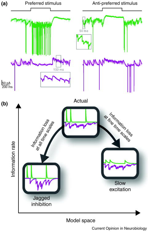
Figure 3.
Shapes of synaptic inputs and relay of information. (a) Membrane current of a relay cell (green) and an interneuron (purple) in response to preferred (left) and anti-preferred (right) stimuli in the center of the receptive field. Insets show magnified synaptic events in time windows marked on the traces. Excitatory (green) and inhibitory (purple) inputs to a relay cell have distinctive shapes: excitation is composed of fast, jagged changes in conductance whereas inhibition is smooth in time. (b) The relayed information about the stimulus is optimized with this synaptic configuration: if excitation is made slower, information at fine temporal resolution will be lost; if inhibition is made jagged, transmitted information will suffer loss at all temporal resolutions [15].

