Fig. 4.
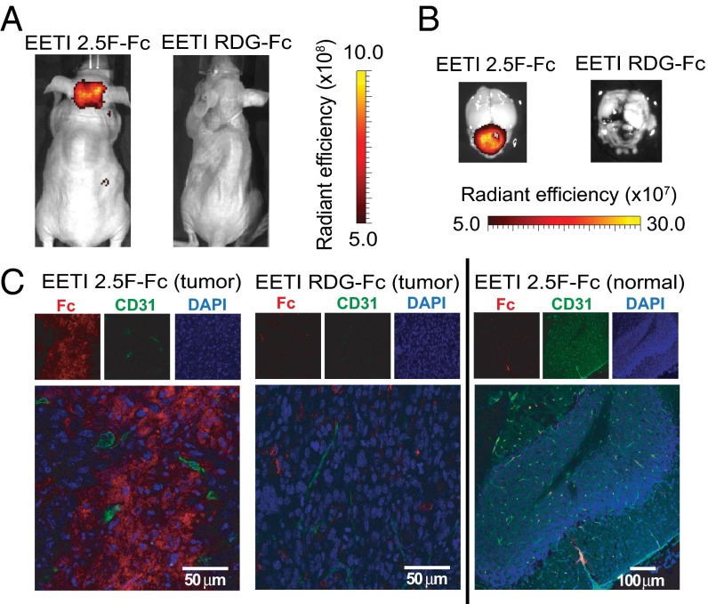
AF680–EETI 2.5F–Fc illuminates MB tumors in vivo and distributes throughout the tumor tissue. Representative in vivo (A) and ex vivo (B) images of signal from tumor-bearing mice injected with AF680–EETI 2.5F–Fc and AF680–EETI RDG–Fc. Images were taken 2 h after probe injection. (C) Brain tissue was processed for histology 2 h postinjection. AF680–EETI 2.5F–Fc was detected with an anti-Fc antibody (red) in tumor tissue costained with the vasculature marker CD31 (green), revealing widespread distribution within the tumor (Left) that was absent in the adjacent normal cerebellum (Right). In contrast, the AF680–EETI RDG–Fc control showed minimal distribution in tumor tissue (Center).

