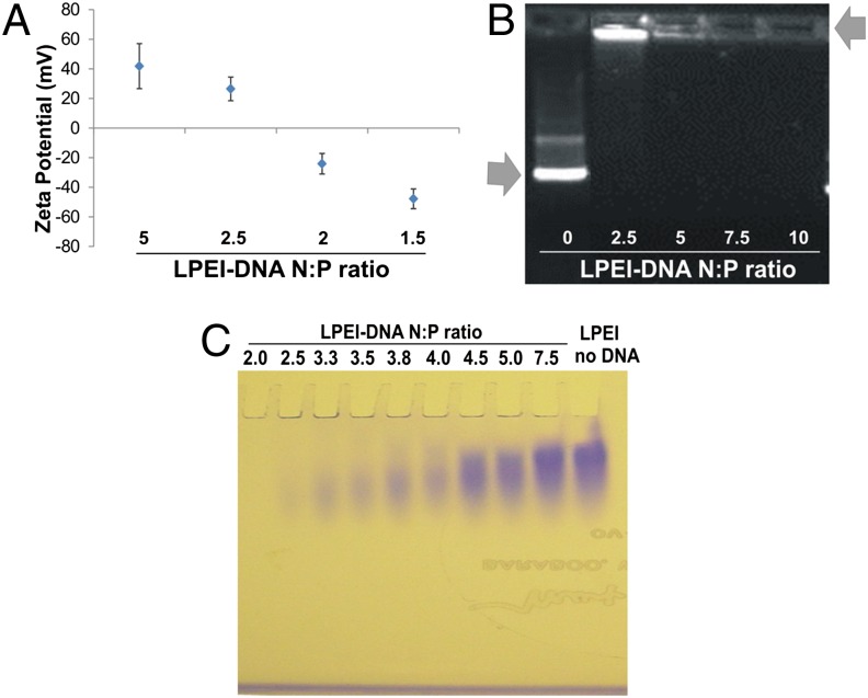
Fig. 1.
Nanoparticle core and shell characterization. (A) Zeta potentials of LPEI88-DNA nanoparticles in double-distilled water (ddH2O) at various N:P ratios. Data represent means and standard deviation (SD) of more than two independent measurements. (B) DNA gel retardation assay. Nanoparticles were formed at various N:P ratios, and electrophoresed at 100 V for 40 min in a 0.5% agarose gel. Arrows point to free DNA and LPEI-complexed DNA. (C) PEI gel electrophoresis of LPEI88-DNA nanoparticles at various N:P ratios and free LPEI88. The LPEI that is not tightly bound to the DNA migrates into the gel and is stained, while the LPEI that is bound to the DNA remains in the well.

