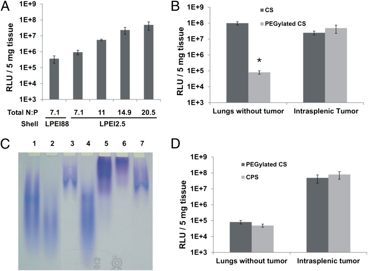
Fig. 2.
CPS formulation. (A) Relative Light Units (RLUs) generated by RLuc (RLuc RLUs) in intrasplenic tumors after transfection with nanoparticles containing LPEI88 as the shell or increasing amounts of LPEI2.5 as the shell. (B) RLuc RLUs in lungs or intrasplenic tumors after treatment with CS nanoparticles or PEGylated CS nanoparticles. (C) Gel electrophoretic analysis of PEGylation of nanoparticles generated with LPEI88 in both core and shell. Lane 1, free non-PEGylated LPEI; lane 2, LPEI purified from the shell of non-PEGylated nanoparticles; lane 3, LPEI purified from the core of non-PEGylated nanoparticles; note that the larger polymer molecules in the heterodisperse LPEI preferentially interacted with DNA and were therefore found in the core; lane 4, free LPEI incubated with non-reactive PEG as a control; lane 5, PEGylated free LPEI; lane 6, LPEI purified from the shell of PEGylated nanoparticles; lane 7, LPEI purified from the core of PEGylated nanoparticles (D) RLuc RLUs in lungs and intrasplenic tumors after transfection with PEGylated CS or CPS nanoparticles. Means and standard deviations of data collected from at least two mice per group are illustrated, * P <0.05.

