Fig. 6.
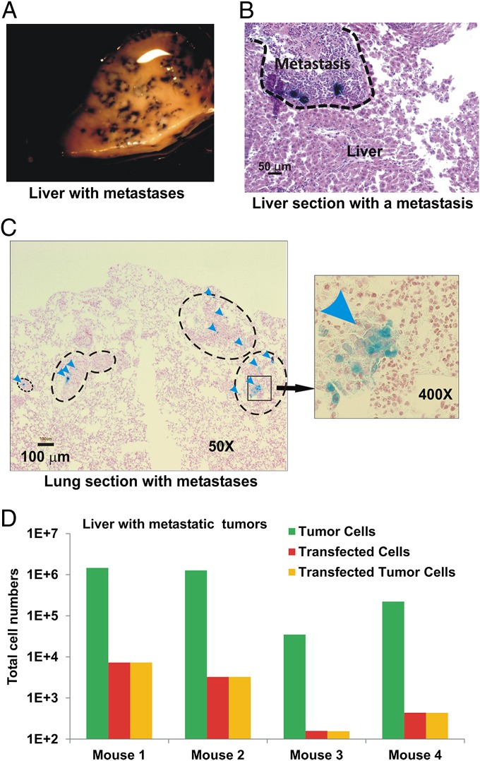
Transfection of metastases with CPS nanoparticles. NOG mice bearing HCT116-Luc2 (A–C) or HCT116-EGFP (D) tumors were transfected with CPS containing β-galactosidase (A–C) or RFP (D) plasmids. (A) Selected view of the transfected metastases in liver. The dark-blue X-gal product accumulates in transfected regions. (B) H&E-stained section of the transfected liver containing a cluster of the transfected metastases. Dark-blue X-gal product can be seen in the transfected tumor cells. (C) Nuclear fast red-stained section of lung containing metastases. Representative metastases and transfected cells (arrowheads) are indicated. (D) Numbers of tumor cells, transfected cells, and transfected tumor cells in the livers with metastases from four individual mice as determined by confocal scanning of dissociated cells.

