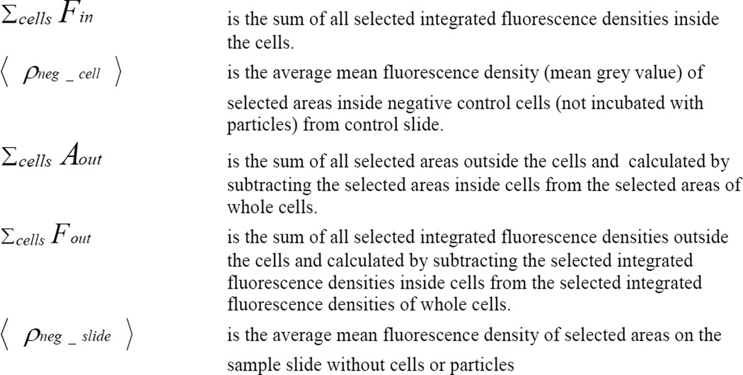
Figure 4. Calculation of fi (internalized fraction of associated particles).
Validation of the method was carried out with fluorescent microbeads. Using 1-micron-sized particles we visually determined and counted the particles inside and outside of cells, and then compared to the data obtained by using the method described here. Figure 5 shows good correlation between the two methods, if one or more microscopic fields were counted.


