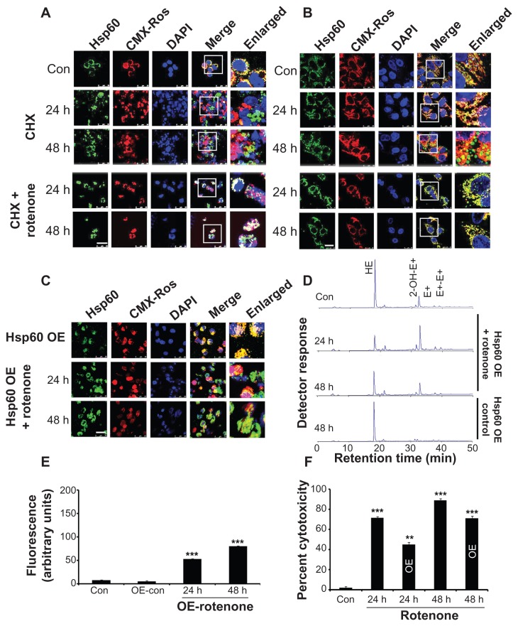
Figure 8.
Comparison of cycloheximide with rotenone-induced Hsp60 translocation. (A) BC-8 and (B) IMR-32 cells were either treated with cycloheximide or combined with rotenone for 24 and 48 h were subjected to cytoimmunofluorescence analysis with anti-Hsp60 antibody (green). (C) BC-8 cells transfected with Hsp60 expressing plasmid either alone or combined with rotenone treatment and for 24 and 48 h and analyzed by cytoimmunofluoresecnce using the anti-Hsp60 antibody. Mitochondria were stained with CMX-Ros (red) and nucleus with DAPI (blue). Images were captured at 63× with a scale bar 25 μm. (D) Analysis of HE oxidation products by HPLC in Hsp60-transfected cells either alone or in combination with rotenone. (E) Analysis of AmplexRed oxidation product, resorufin by spectrofluorimetry in Hsp60 transfected cells, either alone or in combination with rotenone. (F) Cytotoxicity assay of control and rotenone treated cells. Experiments (D–F) were repeated at least twelve times and average arbitrary values (mean ± SEM) are presented. **P < 0.01; ***P < 0.001.

