
Figure 2. Detection of P. citri CHS1 and V-ATPase mRNAs (A) and accumulation of RNAi-induced siRNAs (B) in N. benthamiana plants after TMV-CHS1 inoculation.
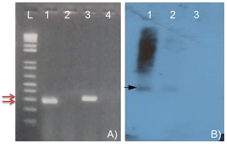
Plants were inoculated with recombinant TMV containing P. citri CHS1 (TMV-CHS1-S) and V-ATPase (TMV-V-ATPase-S). At 7 days post inoculation, total and small RNAs were isolated from infected plants and analyzed by RT-PCR and siRNA northern blot hybridization, respectively. A) One step RT-PCR was performed using total RNA as a template and CHS1 and V-ATPase primers and products analyzed on the gel. Lane L: 1Kb plus DNA ladder, Lane 1: CHS1 primers and RNA of TMV-CHS1-S inoculated N. benthamiana plants; Lane 2: CHS1 primers and RNA of TMV inoculated plant; Lane 3: V-ATPase primers and RNA of TMV-V-ATPase-S inoculated plant and Lane 4: V-ATPase primers and RNA of TMV inoculated plant. B) Small RNAs were separated by PAGE in 15% acrylamide, 8 M urea gels and processed for northern blot hybridization using 32P-UTP-labeled negative strand P.citri CHS1 transcript as a probe. Lane 1: TMV-CHS1-S inoculated plant; Lane 2: TMV-CHS1-AS iinoculated plant; Lane 3: TMV-inoculated plant.