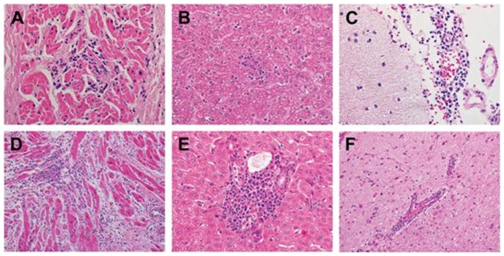
Figure 2. Postmortem histopathologic findings (hematoxylin-eosin (H&E) stain) of two cynomolgus monkeys injected with 107 pfu of CVB3-H3.
Myocardium from both FP42 (A (200X)) and FP49 (D (200X)) showed evidence of viral myocarditis, consisting of focal predominantly lymphocytic mononuclear inflammatory infiltrates with myocyte injury. In addition, representative sections of the liver from both animals exhibited changes of mild viral hepatitis including focal mononuclear inflammatory infiltrates within the hepatic lobules (B (FP42)) and portal tracts with associated interface hepatitis (E (FP49)). The central nervous system exhibited perivascular lymphocytic infiltrates within the leptomeninges (C (FP42)) and intracerebral blood vessels (F (FP49)) with microglial proliferation and nodules (not shown). These changes are indicative of viral meningitis and encephalitis (meningoencephalitis).

