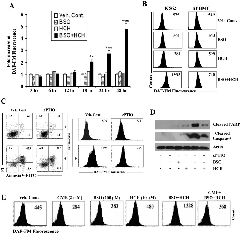
Figure 5. Combination of BSO and HCH induces NO production in CML cells that causes apoptosis.
(A) K562 cells were treated as indicated for different time periods and then intracellular nitric oxide (NO) was measured by flow cytometry after staining with DAF-FM. Data represent mean ± SD of three experiments. ** p<0.01 compared to treatment with either BSO or HCH alone. *** p<0.001 compared to treatment with either BSO or HCH alone. (B) Representative histograms of DAF-FM staining in K562 cells and hPBMC after treatment as indicated for 24 h. (C) K562 cells were pretreated with 100 µM cPTIO for 2 h followed by incubation with combination of 100 µM BSO and 10 µM HCH for 24 h. Cells were then subjected to annexin V/PI binding assay by flow cytometry (left panel). Histograms show measurement of NO in the presence or absence of cPTIO after indicated treatment (right panel). Representative of two similar experiments. (D) K562 cells were treated as indicated for 24 hours after pretreatment with 100 µM cPTIO for 2 h. The whole cell lysates were then immunoblotted with indicated antibodies. (E) K562 cells were pre-incubated with indicated concentrations of GME followed by treatment with BSO and HCH as indicated for 24 h. Analysis of intracellular NO was done by flow cytometry after staining with DAF-FM. Histograms are representative of two similar experiments.

