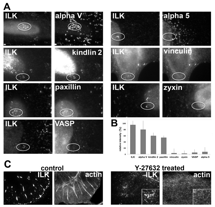
Figure 6. Molecular composition of the novel central adhesions formed following Rho-kinase inhibition.
(A) HeLa JW cells were treated with 10 µM Y-27632 for 60 min, and double-labeled for ILK, the most prominent component of these adhesions, as well as for additional FA components. The same field is shown for two labeled proteins; circles indicate the region of interest. (B) Relative intensity of the novel central adhesions marked by the studied proteins, following 60 min of Y-27632 treatment. The relative intensity was calculated as the percentage of fluorescence intensity measured for the corresponding protein in the untreated FAs. (C) Comparison of ILK and actin localization in FAs of untreated cells, and in the central adhesions of Y-27632 treated cells. Two-color TIRF microscopy of the cells prior to (left panel) and following 60 min of Y-27632 treatment (right panel) was used to assess the localization of ILK and actin. No correlation was found between the densities of these two proteins in the central adhesions (see inserts).

