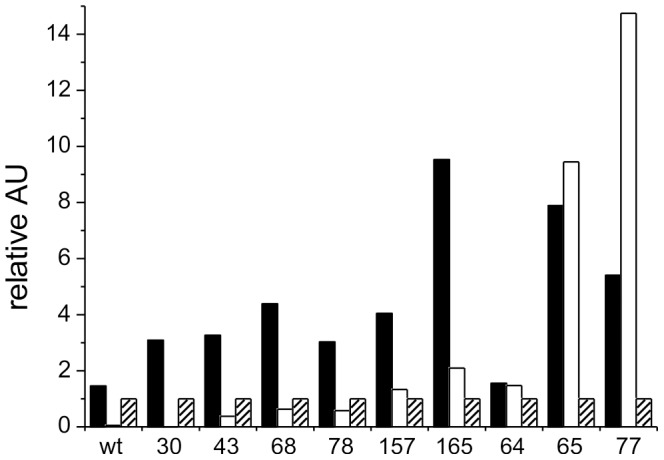
Figure 5. Antiphage ELISA readout values of representative phage clones selected from a 12-mer phage display peptide library.

Solid bars indicate binding of the phage clone to bR loaded nanodiscs, open and hatched bars to wells of the micro titer plate that were loaded with empty nanodiscs or BSA, respectively. Readout values of the wells loaded with bR and empty nanodiscs were normalized to the well binding signal of each clone. Single determination was performed for each clone. Numbers below the bars refer to the given ID numbers of each phage clone. Wild type (wt) refers to phage displaying no peptide.
