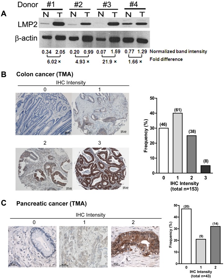
Figure 1. β1i is frequently expressed in colorectal and pancreatic cancer tissues.
(a) Immunoblotting analysis for β1i using protein lysates prepared from nonmalignant (N) and cancerous (T) colonic tissues from the same donors (n = 4 pairs). β-actin was used as a loading control. The band intensities for β1i and β-actin were densitometrically analyzed and used to obtain the relative β1i expression normalized to β-actin levels. (b) Immunohistochemical staining for β1i using a tumor array containing 153 evaluable tumor colon tissue specimens. The intensities of β1i positive staining were evaluated on a scale of 0 to 3. Approximately 70% (46 specimens had intensity of grade 1, 38 with grade 2, and 8 with grade 3, out of 153 total tumor specimens) of colorectal tissues have positive (the staining intensity ≥1) β1i staining. (c) Immunohistochemical staining for β1i using a tumor array containing 43 evaluable tumor pancreatic tissue specimens. The intensities of β1i positive staining were evaluated on a scale of 0 to 2. Approximately 53% (9 specimens with intensity grade 1, and 14 with grade 2, out of 43 total tumor specimens) of pancreatic cancer tissues had ≥1 β1i staining intensity.

