
Figure 3. Representative T1-weighted image (T1WI), T2-weighted image (T2WI), T2*-weighted image (T2*WI) and susceptibility-weighted imaging (SWI) of a 42 year-old patient with splenic siderotic nodules.
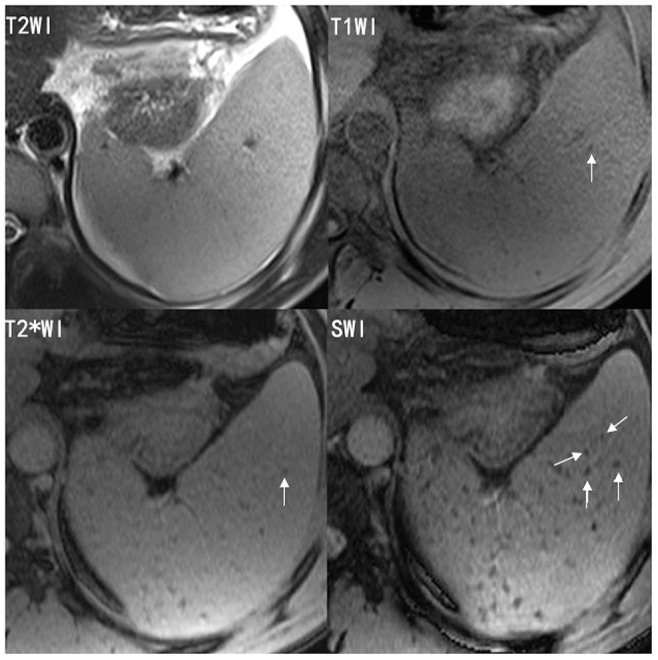
SWI shows more siderotic nodules than T1WI, T2WI or T2*WI.