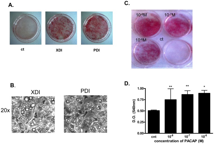
Figure 1. Bright field and Oil-Red-O staining photographs of 3T3-L1 cells.
A. 2 days post-confluent cells were induced to differentiate by treatment with 500 µM IBMX, 0.25 µM dexamethasone and 10 µg/ml insulin (XDI cocktail) or 10−7 M PACAP, 0.25 µM dexamethasone and 10 µg/ml insulin (PDI cocktail), as described under Material and Methods. The cells were stained with Oil-Red-O and photographed on day 9. B. Micrographs of 3T3-L1 cells induced to differentiate with IBMX, dexamethasone and insulin (XDI) and PACAP, dexamethasone and insulin (PDI). C. Oil-Red-O staining of cells induced to differentiate with increasing concentrations of PACAP (10−8 M to 10−6 M), 0.25 mM dexamethasone and 10 µg/ml insulin. Control cells were maintained in medium without induction with hormones. D. Triglyceride content in cells induced to differentiate with increasing concentrations of PACAP (10−8 M to 10−6 M), 0.25 mM dexamethasone and 10 µg/ml insulin. Data were analyzed using repeated measure of ANOVA and by Dunnett’s comparison tests. *p<0.05 compared to control.

