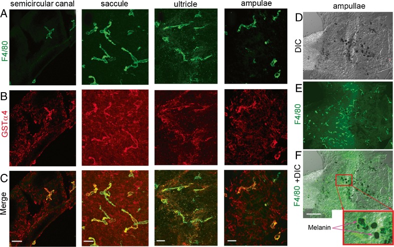
FIG. 2.
Perivascular-resident cells identified as melanocyte-like macrophages. A–C PVM/Ms double labeled with antibody for F4/80 and GSTα4 show positive staining for macrophage and melanocyte marker proteins F4/80 (green) and GSTα4 (red). The scale bar is 50 μm. D–F PVM/Ms contain melanin pigment granules. The column-reticular square region in panel F shows melanin granules under a high magnification. The scale bar is 100 μm.

