FIG. 3.
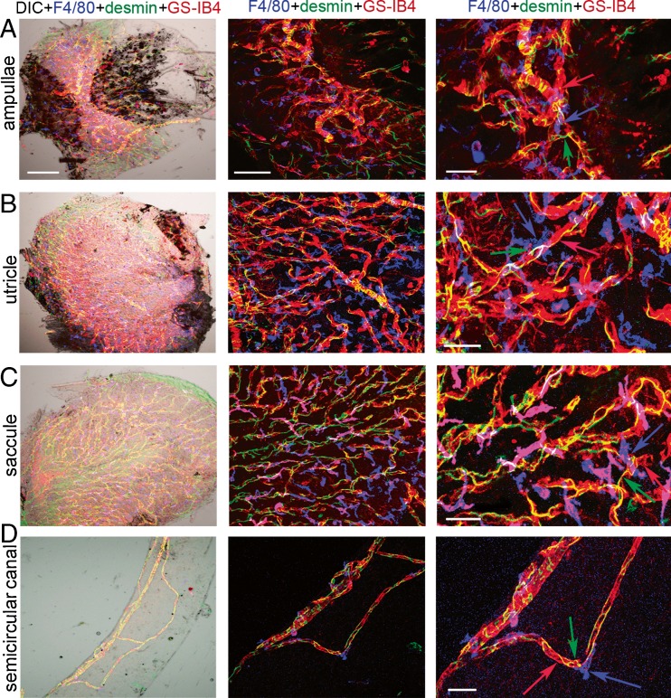
PVM/Ms in the vestibular system are closely associated with microvessels and structurally intertwined with endothelial cells and pericytes. A–D (left panels) Whole-mounted semicircular canal ampullae, utricle, saccule, and semicircular canal triple labeled for F4/80 (blue), desmin (green), and GS-IB4 (red) in low magnification (×20) confocal and DIC images show PVM/Ms, pericytes, and endothelial cells. The scale bar is 100 μm. A–D (middle panels) The confocal image under higher magnification (×40) shows the interaction between the three cell types. The scale bar is 50 μm. A–D (right panels) The zoomed image shows the three cell types to structurally intertwine. The scale bar is 30 μm.

