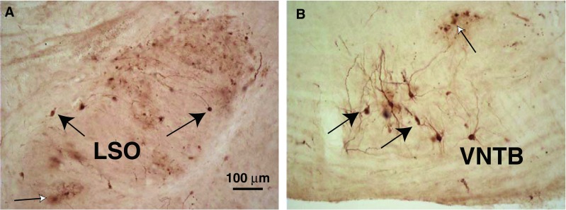
FIG. 1.

Photomicrographs of PRV-labeled neurons. A Labeled LOC neurons (black arrows) in the lateral limb of the lateral superior olive (LSO) on the side ipsilateral to the injected cochlea after a 3-day survival time. “Cloudy” areas of reaction product (white arrow) were sometimes observed in areas of PRV labeling (see text). This section is at the mid-point of the LSO’s rostro-caudal extent. B Labeled MOC neurons (black arrows) in the ventral nucleus of the trapezoid body (VNTB) on the side contralateral to the injected cochlea, in an animal that survived for 3 days after the injection (a different case from A). A labeled group of “blebs” is indicated by a white arrow (see text). This section is from the caudal VNTB, near the caudal tip of the LSO.
