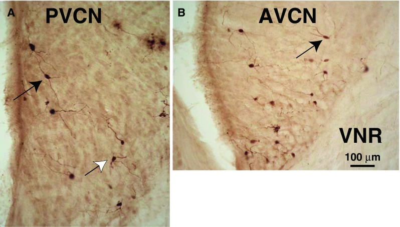
FIG. 7.

Photomicrographs of PRV-labeled multipolar neurons in PVCN (A) and AVCN (B). Planar multipolar cells (black arrows) have long dendrites mostly contained in a single plane and cell bodies of medium size (see text for measurements). Other multipolar cells have more radiating dendrites (white arrow). Both A and B are from the CN ipsilateral to the injection and both had a 3-day survival time. VNR vestibular nerve root. These sections are from a point about midway in the total rostro-caudal extent of the PVCN and AVCN.
