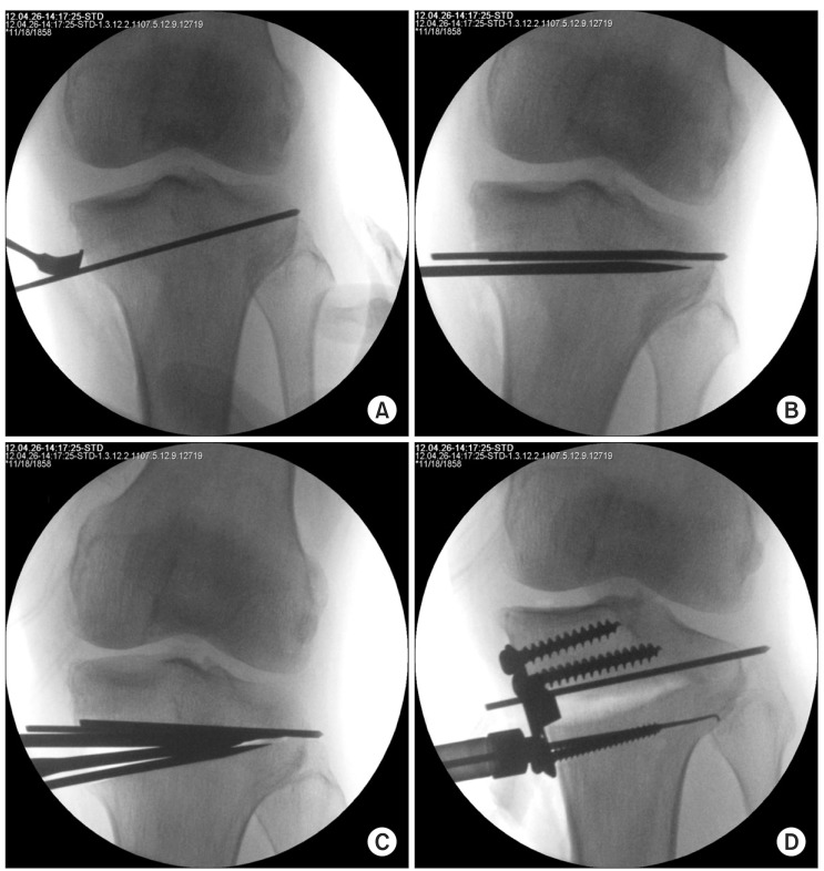
Fig. 1.
Surgical technique. (A) A guide pin was placed from the superomedial portion of the tibial tuberosity to the fibular head approximately 1 cm below the lateral articular margin of the tibia. (B) Osteotomy was advanced with an osteotome to 5 mm medial to the lateral cortex. (C) Two osteotomes were placed in the osteotomy site to spread the osteotomy site without collapsing cutting surface and making intra-articular fracture and lateral hinge tears. (D) The medial tibia was fixed with a Puddu plate.

