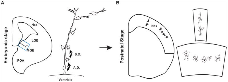
Figure 5. Clonal production and organization of inhibitory interneurons in the mouse neocortex.

(A) At embryonic stage, RGCs in the VZ of the MGE divide asymmetrically to self-renew and to simultaneously produce differentiating interneurons or IPCs that divide symmetrically in the SVZ to produce differentiating interneurons. The progeny of the same RGC initially migrate along the mother RGC and form radially aligned clonal clusters (the clone in the MGE is shown at higher magnification in the right panel). The new-born cells progressively move away from the VZ, differentiate, and migrate tangentially towards the neocortex. (B) After arriving at their destination in the neocortex, inhibitory interneuron clones do not randomly disperse, but form spatially organized clusters which are either vertically or horizontally oriented (one example in each case is shown at higher magnification in the right panel). A.D., asymmetric division; S.D., symmetric division. Adapted from Fig. S14 114.
