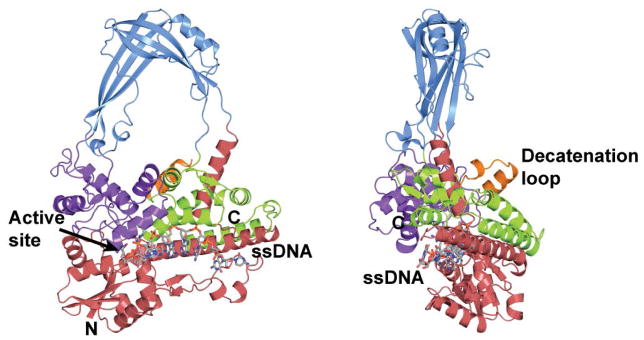
Figure 1. Structure of E. coli topoisomerase III, a member of the type IA family of topoisomerases.

The diagram shows the structure of the intact E. coli topoisomerase III in complex with single stranded DNA (15). The enzyme is formed by four domains (red, blue, purple, and green) that form a toroidal shape with a central hole. The active site is found at the interface of two of the domains. Single stranded DNA binds along a groove before it enters the active site. Topoisomerase III has a 17 amino acid loop, the decatenation loop, which is needed for decatenation activity (32). Topoisomerase I does not have this loop.
