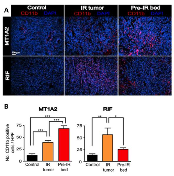
Figure 2. Irradiation produces influx of bone marrow derived CD11b+ monocytes into tumors.
(A) Immunostaining for CD11b (red) in tumors with no IR (control), irradiated tumors (IR tumor), or tumors grown in the irradiated bed (pre-IR bed) for MT1A2 (upper panel) and RIF (lower panel). Nucleus staining with DAPI is shown in blue. (B) Quantification of CD11b+ myelomonocytic cells in (B) for MT1A2 (left) and RIF (right) tumors. Symbols and error bars represent the mean ± SEM for n>4 animals per group. *p < 0.05, **p < 0.01, and ***p < 0.001, respectively, determined by one-way ANOVA. From (13) with permission.

