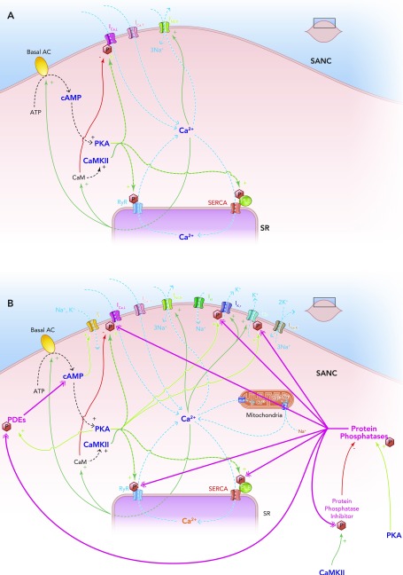
FIGURE 5.
The coupled clock system
A: Ca2+ release begets Ca2+ release. A stripped down schematic figure illustrating how, without highly constitutively active phosphodiesterases and protein phosphatases, the coupled clock system would inexorably accelerate itself toward its fastest rate. B: schematic demonstration of the sites of action of the highly constitutively active phosphodiesterases (PDEs) and protein phosphatases (PPs) that keep the coupled clock system in check, allowing it to operate approximately in the mid-range of its capabilities. Note that protein kinase A phosphorylates and activates PDEs, increasing their activity; this is antagonized by the activity of PPs. Protein kinase A also activates PPs directly. Ca2+-calmodulin kinase II phosphorylates and activates protein phosphatase inhibitors, which go on to inhibit PPs, which in turn antagonize the phosphorylating effect of CAMKII on PP inhibitors.

