
FIGURE 3.
Work on diving rodents suggest paranasal areas (shaded blue) innervated by the anterior ethmoidal and infraorbital nerves are important for initiating the diving response
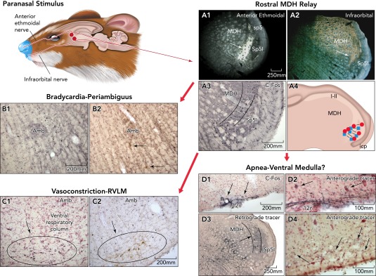
These nerves project [A1 and A2 show transport of an HRP cocktail (colored gold) transported transganglionically from the anterior ethmoidal and infraorbital nerves, respectively] into the rostral medullary dorsal horn (MDH) overlapping the caudal subnucleus interpolaris (Sp5I). Note the band of neuropil just dorsal to the Sp5I (arrows) is labeled from either nerve. Neurons activated with cFos (A3; small black nuclei) induced by diving are found in similar neuropil. Moreover, small, bilateral injections of lidocaine (blue squares) or kynurenate (red circles) made into similar locations (A4) blocked the cardiorespiratory responses of nasal stimulation. The hallmark of the diving response is the dramatic bradycardia (see FIGURE 1A); many neurons surrounding the rostral nucleus ambiguus are labeled with cFos after diving (B1), and some of these are preganglionic cardiac motoneurons (B2; arrows point to double-labeled neurons containing cFos and a retrograde tracer injected into the pericardial sac). There also is a massive but selective peripheral vasoconstriction during diving in rats mediated by neurons in the rostral ventrolateral medulla (C1 shows cFos-labeled neurons in the RVLM induced by diving); many such neurons are monoaminergic (C2 showing double labeled neurons with antibodies against cFos and tyrosine hydroxylase). The third neuronal reflex induced by underwater submergence is a profound apnea, which is maintained despite gross disruption of blood chemistry, suggesting inhibition of the respiratory chemoreceptor reflex. Few neurons were activated in the medullary ventral respiratory column (see C1, C2), but projections from the MDH to the ventral surface of the caudal medulla at the spinomedullary junction (D2; approximately −14.6 mm from bregma) overlap where neurons/glia activated by diving are found (D1, arrows; small black profiles show cFos activation). Arrows in D2 point to presumptive neurons with juxtaposed BDA fibers. Injection of a retrograde tracer, which included the retrotrapezoid nucleus labeled small neurons in neuropil similar to that labeled by paranasal primary afferent fibers (D3, arrow). Anterograde transport of tracers injected into these areas of the MDH resulted in extremely small labeled fibers with swellings (D4, arrows) in the retrotrapzoid nucleus ventral to the facial motor nucleus. Similar neurons/glia have long been suspected to be chemoreceptors sensitive to high Pco2, but details of how they interact with central respiratory neurons is lacking. Other studies (188) have shown the neuronal circuitry driving the diving response is contained within the medulla and spinal cord.