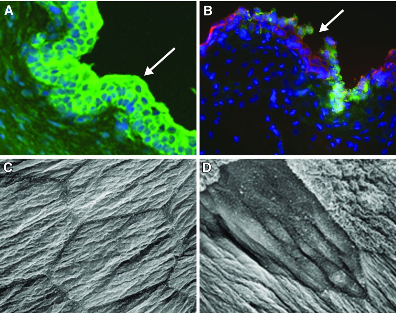
Figure 6.
Urothelial alterations following injury or inflammation. A: intact urothelium in rat urinary bladder (green pan-cytokeratin labeling epithelium; blue, DAP-I nuclear marker). B: area of damage (shown at arrow) to the rat urothelium following cyclophosphamide treatment in a rat. (Modified from Birder 2012.) C and D: scanning electron micrograph of apical surface of umbrella cell layer from normal rat (C) and 2 h after spinal cord injury (D). [Modified from Apodaca et al. (22).]

