Figure 1.
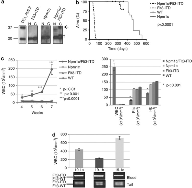
Npm1c and Flt3-ITD collaborate to drive rapid-onset leukemogenesis with frequent occurrence of Flt3 LOH. (a) Npm1 mutant protein (arrow) accumulates in the cytoplasm of spleen cells collected from 3-week-old Npm1c/Flt3-ITD, but not Npm1c or Flt3-ITD single-mutant mice. (b) Kaplan-Mayer survival plots showing the rapid demise of Npm1c/Flt3-ITD mice compared with all other genotypes. (c) Serial blood counts highlight a consistent explosive increase in blood leukocytes counts (WBC) between 4 and 7 weeks in Npm1c/Flt3-ITD mice (left) and the markedly abnormal WBC, platelet count (Plts) and hemoglobin concentration (Hb) of sick leukemic Npm1c/Flt3-ITD mice compared with age-matched control mice. (d) Loss of the Flt3 WT allele in blood DNA from Npm1c/Flt3-ITD AMLs is demonstrated as loss of intensity of the Flt3-WT PCR band. By contrast, constitutional tail DNA shows no LOH. In these three littermates (19.1a–c), the extent of Flt3-LOH associates with the degree of leukocytosis (N=nuclear lysate, C=cytoplasmic lysate, OCI-AML3 lysate as positive control, † nonspecific band).

