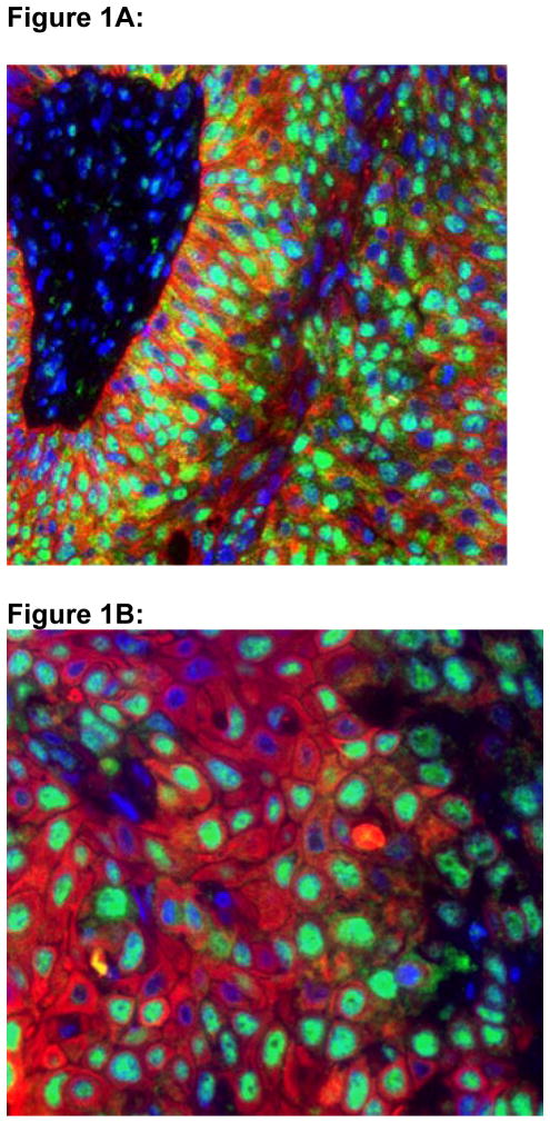
Figure 1.
Figures 1A and 1B: Confocal Microscopy of RRM1 Expression in a Radical Cystectomy Specimen.
RRM1 displays a coarse granular staining pattern with predominant nuclear localization. DAPI staining of nuclei is shown in blue, immunofluorescent staining of cytokeratin is shown in red, and immunofluorescent staining of RRM1 is shown in green. Figure 1A is at 400x magnification; Figure 1B is at 600x magnification.

