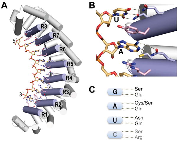
Fig. 1.
PUF domain RNA interaction scaffold. A. Ribbon diagram of a crystal structure of human Pumilio 1 RNA-binding domain in complex with RNA ligand (5′-UGUACAUA). RNA interaction helices are shown as light blue cylinders and labeled R1–R8. Edge-interacting side chains from each repeat are colored light blue, stacking side chains are colored pink, and RNA is colored peach. For stick representations, nitrogen atoms are blue, oxygen atoms are red, and sulfur atoms are yellow. B. Interaction of PUF repeats with uracil or adenine bases. Hydrogen bonds or van der Waals contacts are indicated by dotted lines. C. PUF RNA interaction code. Edge-interacting side chains that specify base recognition are shown. G, A, and U were derived from natural PUF proteins while C recognition was selected by screening.

