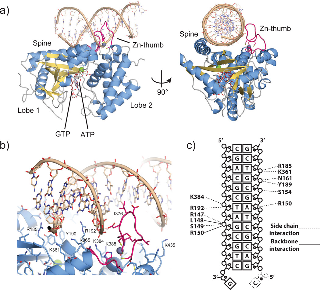
Figure 2. The cGASMab21:DNA:GTP:ATP complex.
a). Side and top views of cGASMab21 (color code of Fig. 1b) in complex with dsDNA (brown), GTP and ATP (ruby stick models). DNA binds along the platform between spine and Zn-thumb.
b) Close up view of the DNA binding site with selected annotated residues. DNA is bound mainly via the minor groove. A notable exception is the Zn-thumb near the major groove.
c) Schematic representation of DNA:cGAS contacts.

