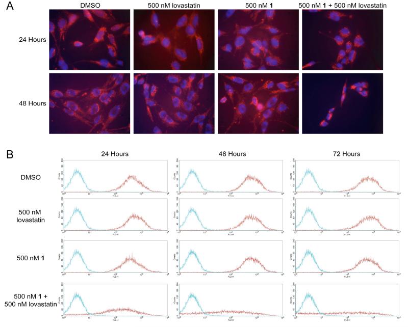
Figure 8.
A, NF90-8 cells were treated as indicated in the figure for 24 and 48 hours. Treated cultures were incubated with 50 nM Mitotracker Orange CM-H2 TM Ros and Hoëchst 33342 to monitor Δψm and nuclear morphology, respectively. Cultures treated with 500 nM 1 in combination with 500 nM lovastatin have reduced Δψm at 48 hours coinciding with chromatin condensation, as compared to the control cultures. B, NF90-8 cells were treated as indicated in the figure for 24, 48, and 72 hours and Δψm was observed using JC-1 by flow cytometry. Histograms represent 2×10⁴ events collected using the FL2-H channel. The red data points denote NF90-8 cells stained with JC-1, and the blue data points denote untreated NF90-8 cells not stained with JC-1. NF90-8 cultures treated with DMSO, 500 nM 1, or 500 nM lovastatin maintained Δψm as shown by the presence of red emission from 24 to 72 hours. However, 500 nM 1 in combination with 500 nM lovastatin resulted in a loss of Δψm.

