
Figure 2. Longitudinal changes in fundus autofluorescence associated with macular atrophy in Stargardt disease in patient #1.
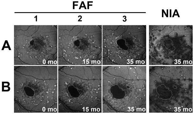
Autofluorescence imaging on FAF and NIA in the right (A) and left (B) eye of a patient with Stargardt disease. Areas of clinically evident macular atrophy have a corresponding area of markedly decreased autofluorescence on FAF imaging; atrophic regions are highlighted (white outline) and can be observed to enlarge by continguous spread with time. The areas of hypoautofluorescence on NIA were larger than the corresponding areas of clnical atrophy and extended beyond the borders of the outlined atrophic areas. Numbers (1,2,3) indicate separate time-points during follow-up.