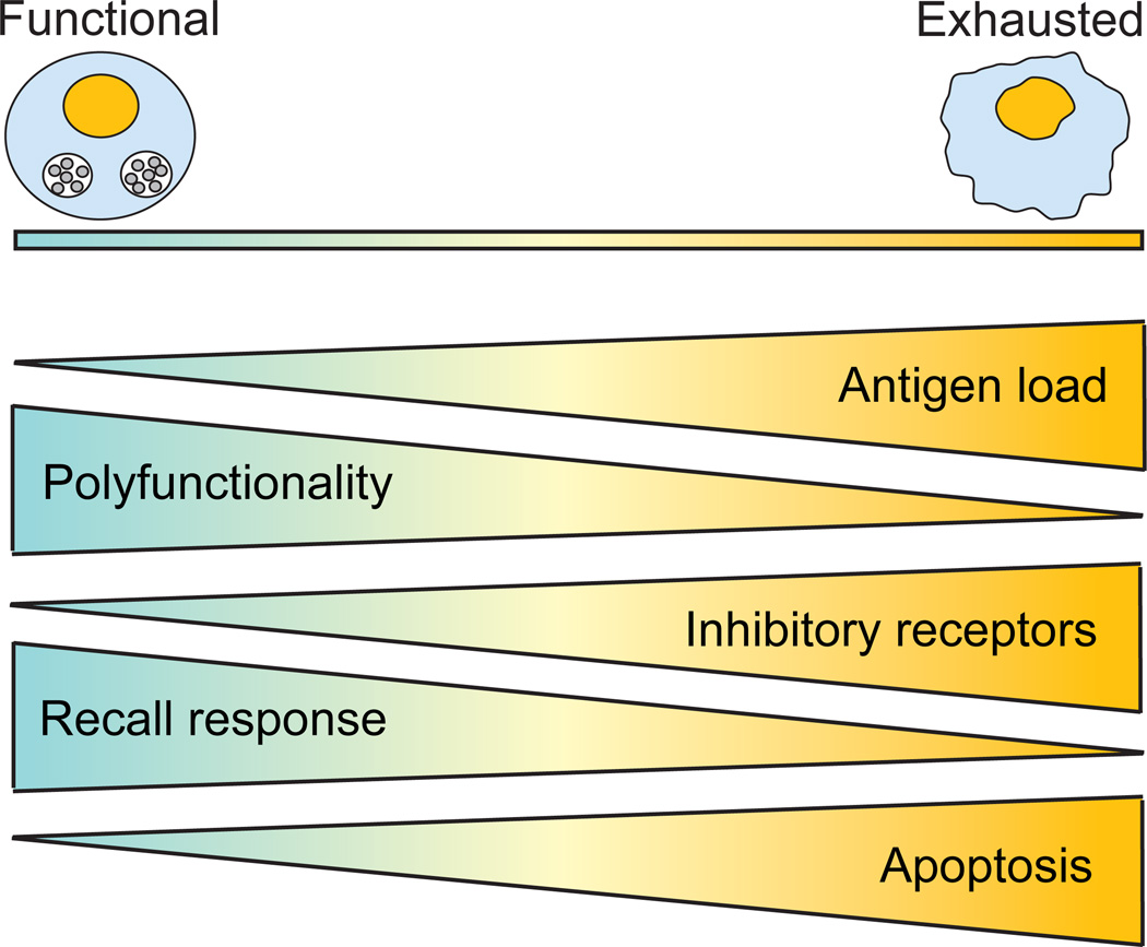
Figure 1. T cell exhaustion.
During acute infections, the host develops a successful T cell immune response against the pathogen, characterized by rapid proliferation and robust polyfunctionality (cytotoxicity and production of IFNγ, TNFα and IL-2). However, during chronic infection, T cells become progressively exhausted and gradually lose the ability to mount an effective recall response to the infection as well as their polyfunctionality ability. At first, reduced IL-2 production and proliferative response are detected. Then, as exhaustion progress, cells lose the ability to produce TNFα. Finally, cells exhibiting the most severe phenotype are unable to secrete IFNγ in response to the infection. At the same time, gradual upregulation of inhibitory receptors (PD-1, LAG3, CD160, CTLA4, 2B4) plays a central role in T cell exhaustion and ultimately, concomitant expression of multiple inhibitory receptors leads to severe T cell exhaustion. Concurrently, exhausted T cells exhibit increased apoptosis potential, leading to their complete deletion. T cell exhaustion is highly dependent on antigen load, and as antigen burden increases, T cells become more exhausted.

