
Figure 1.
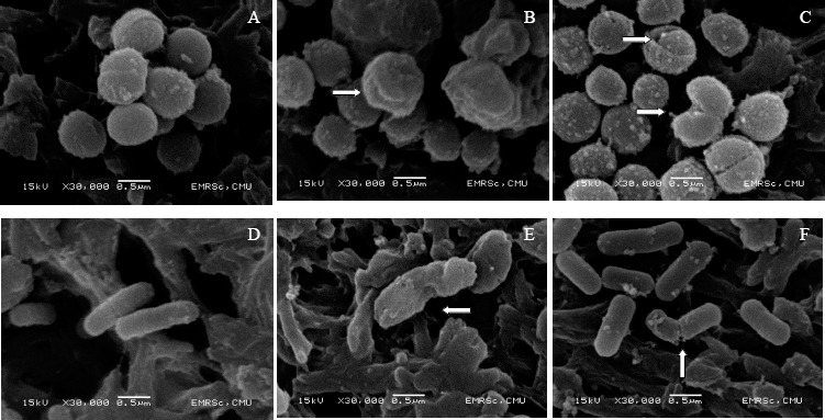
Scanning electron microscope images of S. aureus subsp. aureus DMST 6512 (ATCC 6538Ptm) (left) and L. monocytogenes DMST 17303 (right) cells, in control (MRS broth) (A and D), in the presence of supernatant of L. fermentum FTL 2311 (B and E), and in the presence of L. fermentum FTL 10BR (C and F).