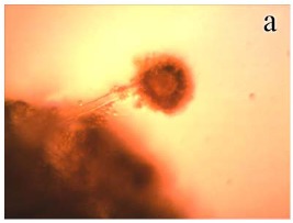
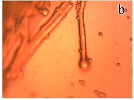
Figure 3.


Light micrographs (X 400) of A. flavus OC1 control (without AGE treatment) and sample (treated with 2.88 mg/ml of AGE). (a): Control conidial head of A. flavus, large and radiated, development of vesicle on conidiophore and conidia are clearly visible; (b): Sample of conidial head of A. flavus showing morphological aberrations induced by AGE. A decrease in conidiation is clearly visible.
