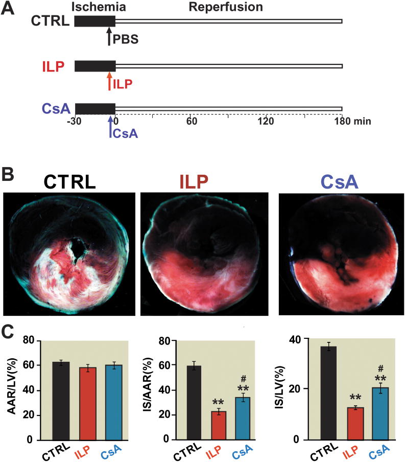
Figure 1. Intralipid reduces the infarct size more efficiently than Cyclosporine-A in the in vivo ischemia/reperfusionrat model.
A. The left coronary artery was occluded for 30 minutes followed by 3 hr of reperfusion. One single IV bolus of 20% Intralipid (5ml/kg body weight, intralipid group), Cyclosporine-A(10mg/kg body weight, cyclosporine-A group) and phosphate buffered saline (control group, CTRL) was administered 5 min before reperfusion. B. representativetriphenyltetrazolium chloride-stained heart slices from from control, intralipid(ILP) and cyclosporine-A(CsA) group. The white area represents show infracted area (Corresponding drawings), blue area shows noneinfarct area red and white areas show risk area. C. Percentage of area at risk (AAR) divided by left ventricle (LV) (B), infarct size (IS) divided by AAR (C), and infarct size (IS) divided by left ventricle in CTRL(n=7), intralipid group(n=7) and cyclosporine-A group (n=7). **p<0.001 vs. CTRL, #p=0.030 vs. CsA.

