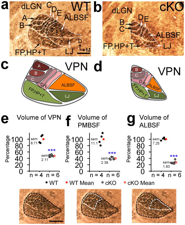
Figure 4. VPN thalamus was re-patterned in cKO mice to match reduced size and in complete body map of S1.
Cytochrome oxidase (COX) staining was performed on PND7 coronal sections to measure VPN volume and to reveal barreloid patterning. (a,b) Compared to wildtype, barreloid row ‘A’ and most ALBSF barreloids were absent in VPN of cKO mice. (c,d)Schematics of the VPN body maps as in a and b. Quantification of VPN body map: (e) VPN volume (47.8% +/− 2.11% of wildtype, p< 0.0001, t = 9.9, df = 8 n = 6), (f) PMBSF barreloid volume (40.08% +/− 2.38% of wildtype, p< 0.0001, t = 8.4, df = 8, n = 6) and (g) ALBSF barreloid volume (28.62% +/− 1.83% of wildtype, p< 0.0001, t = 15.6, df = 8, n = 6). Abbreviations: D: dorsal, VPN: ventroposterior nucleus, dLGN: dorsal lateral geniculate nucleus, A–E: barreloids of whisker rows ‘A’–‘E’, ALBSF: barreloids of anterior snout whiskers, FP, HP+T: barreloids of the paws and rest of the trunk/body, LJ: barreloids of the lower jaw map. Scale bar in a,e, 250μm.

