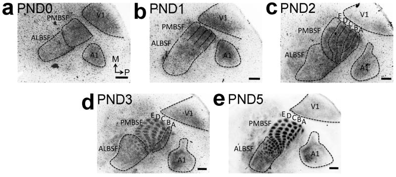
Figure 7. Late differentiation of VPN TCA representations of PMBSF row ‘A’ and ALBSF in S1.

Spatiotemporal progression of the differentiation of body representations formed by TCAs from VPN in the cortical plate of S1 was analyzed by GFP immunohistochemistry on tangential sections of flattened cortex from RORα-IRES-Cre; ROSA26-GAP43-eGFP mice (also see Supplementary Fig. 4). a: At PND0, more GFP-positive TCAs are evident in nascent PMBSF compared to the future ALBSF that is still sparsely innervated by GFP-positive TCAs. b: Although not segregated by septa yet, bands of nascent rostral barrel rows ‘B’, ‘C’, ‘D’ and ‘E’ are evident at PND1 (PMBSF outline b). c: TCAs forming the immature band for nascent caudal row ‘A’ differentiate within the S1 cortical plate significantly later and are first seen at PND2 (outlines of row ‘A’-‘E’ in c). d: By PND3 septa between individual barrels of row ‘B’ to ‘E’ and the later almost ALBSF barrels are formed and fully differentiated. Conversely row ‘A’, still appears as a band (d). e: Individually separated row ‘A’ barrels with differentiated septa are first identifiable later at PND5. Number (n) of brains analysed at each age: PND0: n= 8, PND1: n= 9, PND2: n= 8, PND3: n= 8, PND5: n=7. Scale bar in a–e, 250μm.
