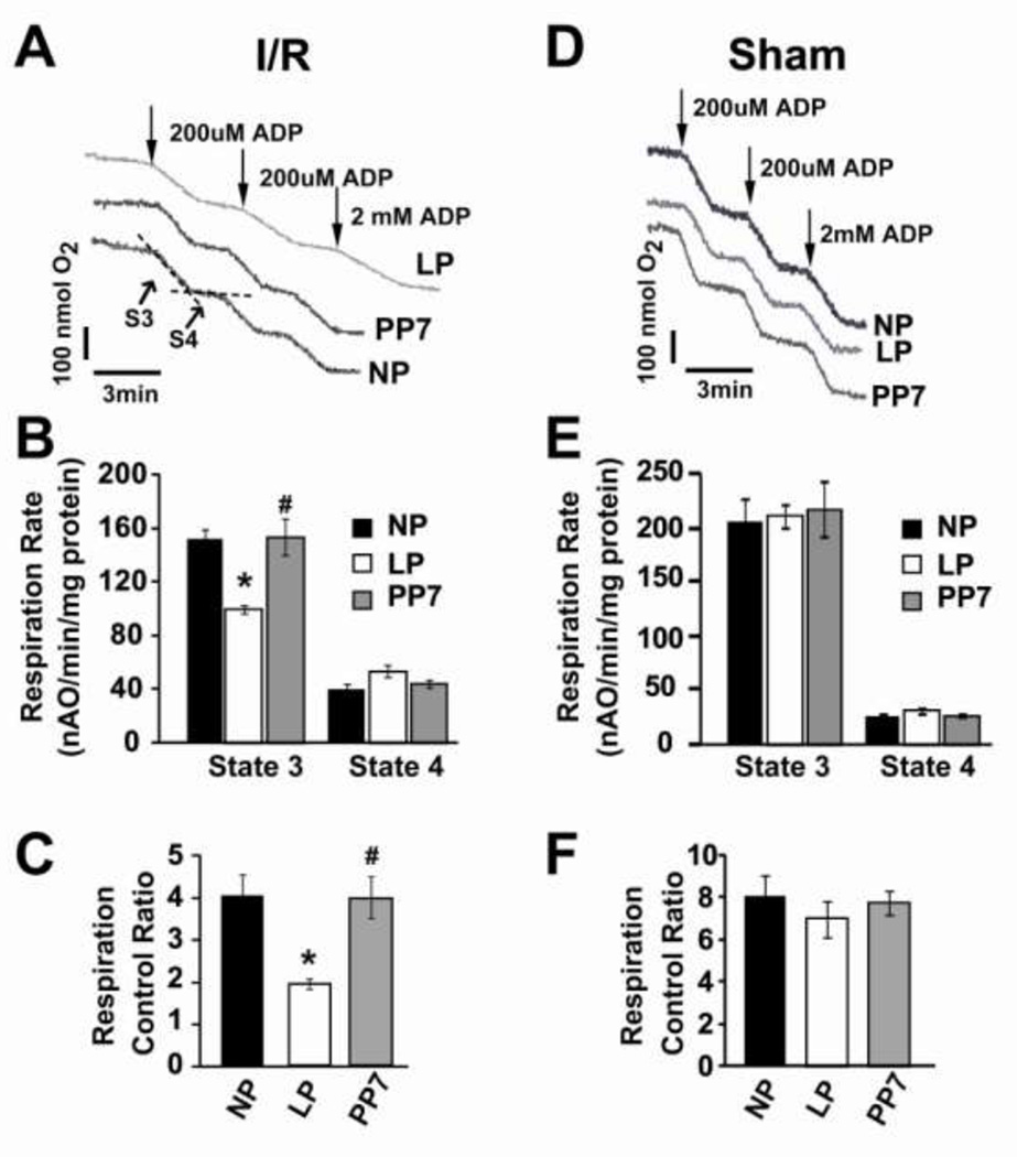
Figure 4. Mitochondrial respiration decreased in LP heart subjected to ischemia reperfusion injury.
A, D. Typical oxygen electrode traces showing the respiration in state 3 stimulated by 0.2 mM ADP (S3) and in resting state 4 (ADP-limited, S4) of complex-I in isolated mitochondria from NP, LP and PP7 subjected ischemia/reperfusion injury (A), or not subjected to ischemia/reperfusion injury (sham, D). B, E. Respiration rate of state 3 and state 4. and C, F. Respiratory control ratio (respiration rate of state 3/state 4) in NP, LP and PP7, **p <0.05 LP vs. NP; #p<0.05 PP7 vs. LP; p >0.05 NP vs. PP7(n=4).

