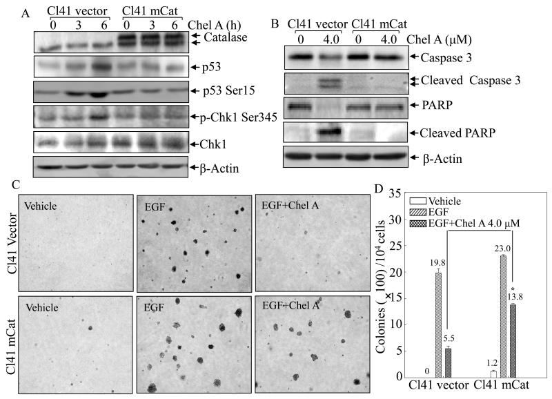
Fig. 6. mCat overexpression blocked the biological effect of Chel A in Cl41 cells.
(A and B) Cl41 mCat and Cl41 vector were treated with Chel A for 0-6 h (A) or 48h (B). The cell extracts were subjected to Western blotting. β-Actin was used as protein loading controls. (C and D) Effect of Chel A on EGF-induced cell transformation in Cl41 mCat cells and Cl41 vector cells were determined by Soft Agar assay. The colony formation was observed under inverted microscope and photographed (C). The numbers of colonies were scored, and presented as colonies per 10,000 seeded cells (D). The symbol (*) indicates a significant increase in Cl41 mCat cells as compared with Cl41 vector(p<0.05). Each bar indicates the mean and standard deviation from three independent experiments.

