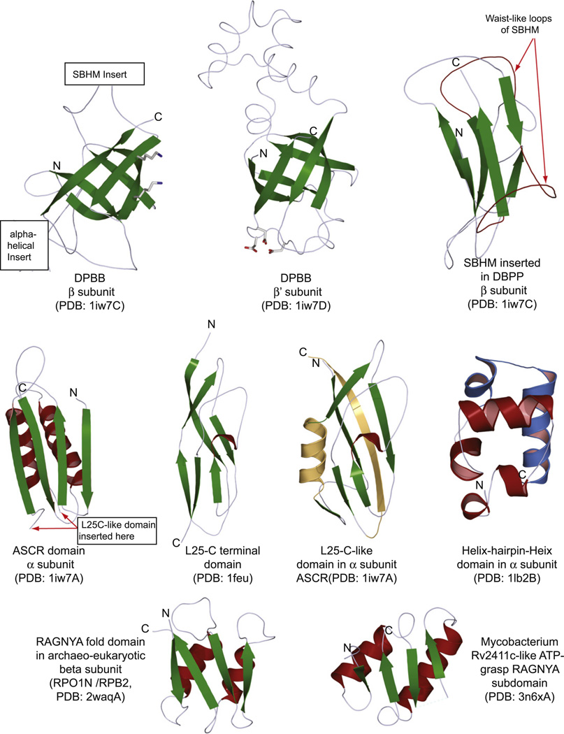
Fig. 2.
Structures of key conserved domains of the β, β′ and α subunits. Strands are colored green, whereas helices are colored red or blue. Only the core conserved regions of the domains are shown. Inserts in domains are mostly suppressed or excised as depicted. The C-terminal domain of the ribosomal L25 protein is also depicted to illustrate its structural relationship with the conserved domain inserted into the ASCR domain of the α subunit (L25C–like domain). Structural elements in the L25C–like domain of the α subunit that are not present in the ribosomal L25 protein are colored orange.

