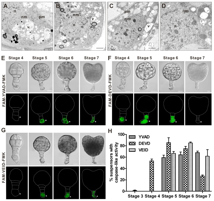
Figure 3. Vacuolization of the cytoplasm, nuclear envelope disassembly, and caspase-like proteolytic activity during suspensor PCD.
(A–D) Morphology of the basal cell analysed by TEM in the embryos at stages 6 (A), 7 (B), 8 (C), and 9 (D). Scale bars, 2 µm in (A–C) and 1 µm in (D). n, nucleus; v, vacuole; nm, nuclear membrane; pm, plasma membrane. (E–G) In situ detection of active proteases with caspase 1-like (E), 3-like (F), and 6-like (G) specificity in the suspensor cells at stages 4 to 7, as revealed by staining with indicated caspase-specific fluorescent inhibitors. Scale bars, 20 µm. Asterisks denote the basal cell. (H) The frequency of suspensors with caspase-like activity at the developmental stages 3 to 7. Data represent the mean ± SE from four independent experiments, with 30 embryos per stage analysed in each experiment (n = 120).

