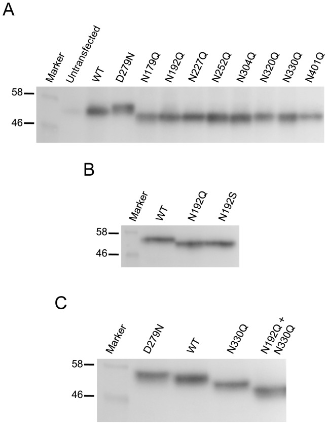
Figure 1. All eight putative N-glycosylation sites of CLN5 are utilized in vivo.
HeLa cells were transiently expressing the various N-glycosylation mutants of CLN5 as indicated. The whole cell lysates were collected 24 h post transfection and analyzed by Western blotting. (A) wt CLN5, D279N, and eight single N-glycosylation site-deleted mutants as indicated. (B) Comparing N192Q and N192S (patient mutation) migration on gel. (C) Comparing migration on gel of single and double N-glycosylation site mutants. Equal amount of lysates was loaded onto each well. The mouse monoclonal anti-Myc antibody was used to detect CLN5.

