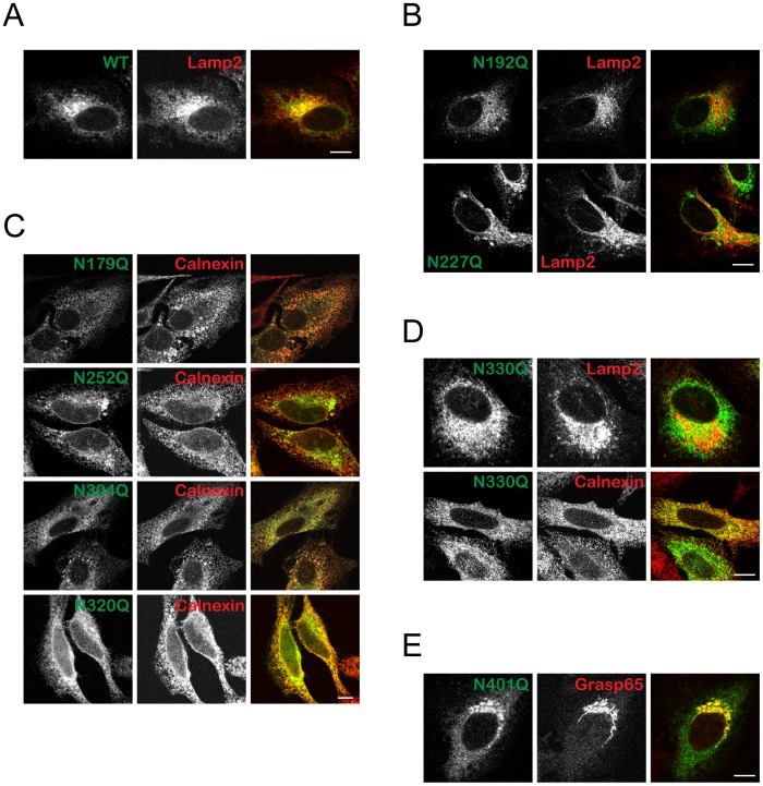
Figure 2. Subcellular localization of CLN5 N-glycosylation mutants.
Confocal microscopy analysis of cells transiently expressing N-glycosylation mutants of CLN5. HeLa cells were seeded on glass coverslips and transfected with wt CLN5 or mutants. The cells were treated with cycloheximide for 2 h prior to fixation. Different antibodies were used to label specific organelles (A), (B) and (D) Lamp2 for the lysosomes, (C) and (D) Calnexin for the ER, (E) Grasp65 for the Golgi. Cln5 mutants are as indicated. The mouse monoclonal anti-Myc antibody was used to detect CLN5. Original magnification, 1,000×. Bars, 5 µm.

