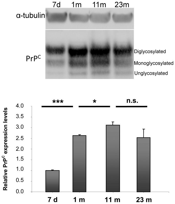
Figure 1. PrPC expression levels in membrane extracts from mouse hippocampi at different developmental stages.
Western blot analysis of equal amounts of protein from hippocampal membrane extracts (25 µg per lane) at the indicated ages. 7 d = 7 days old; 1 m = 1 month old; 11 m = 11 months old; 23 m = 23 months old. Each lane corresponds to a single animal. Antibodies used: D18 (1:1,000; InPro Biotechnology, Inc, South San Francisco), mouse monoclonal anti α-tubulin (1:10,000; Calbiochem). The three major PrPC glycosylation forms are visible. Relative PrPC expression levels were analyzed from 3 to 4 mice per time point. Each data point represents the relative protein level normalized over α-tubulin ± SD. Changes in band intensity were analyzed and quantified with ImageJ 1.37v software (NIH, USA) followed by comparison with ANOVA test for groups of mice at different ages. Differences were considered significant when p<0.05. PrPC levels in hippocampal membrane from mice increased dramatically at the time of synaptogenesis (1 m), rose further during adulthood (11 m) and then remained at plateau during aging (23 m).
n.s.: not significant, *: p<0.05, ***: p<0.001.

