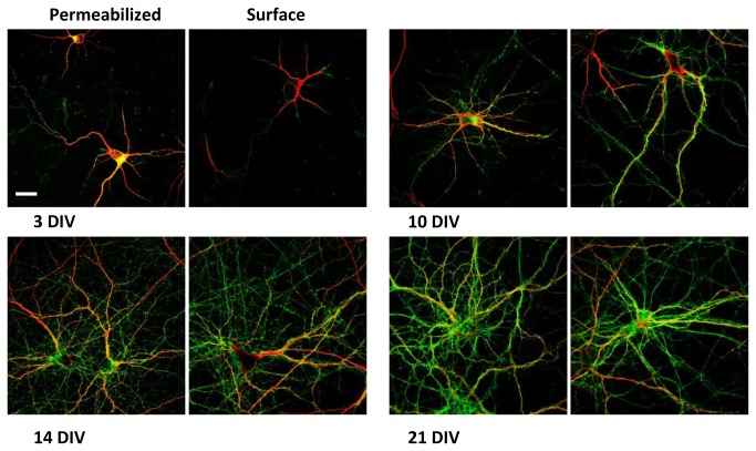
Figure 7. PrPC co-immunolabeling with MAP2.
Confocal images of hippocampal primary neurons at different developmental stages. Normal (left) and surface (right) immunolabeling of PrPC (green) coupled with MAP2 staining (red). Antibodies used: D18 (10 µg/mL and 20 µg/mL in surface immunolabeling InPro Biotechnology, Inc, South San Francisco), rabbit polyclonal anti MAP2 (1:500; Santa Cruz).
Scale bar = 10 µm.

