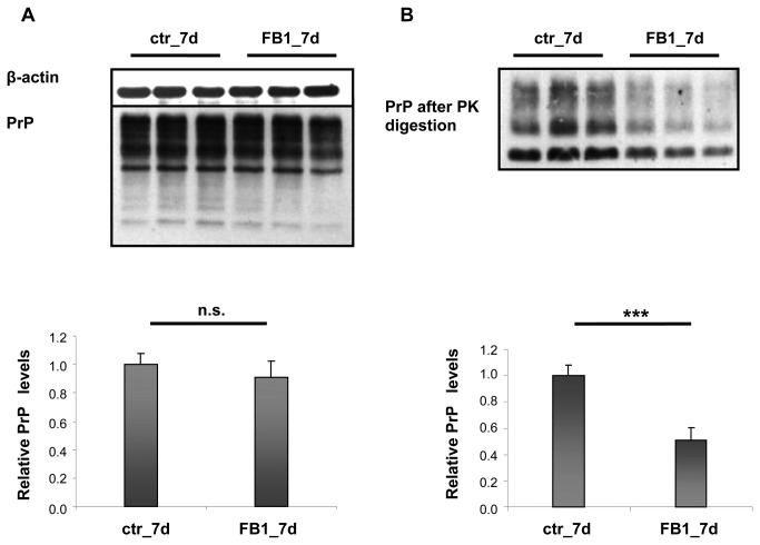
Figure 10. Total PrP and PK-resistant PrP in ScGT1 cells after treatment with FB1.
ScGT1 cells were treated for 7 days (7d) with FB1 (25 µM). A) Western blot analysis of equal amounts of protein from ScGT1 cells (25 µg per lane). Antibodies used: D18 (1:1,000; InPro Biotechnology, Inc, South San Francisco), mouse monoclonal anti β-actin (1:25,000; Sigma-Aldrich). Each data point represents the mean protein level normalized over β-actin ± SD.
B) Western blot analysis of equal amounts of protein from ScGT1 cells (250 µg per lane) after PK digestion. Antibodies used: D18 (1:1,000; InPro Biotechnology, Inc, South San Francisco) and mouse monoclonal anti β-actin (Sigma). Each data point represents the mean protein level normalized over total PrP ± SD. No significant changes in PrP levels were detected in the total protein extracts or in protease-resistant PrP after sphingomyelin treatment. FB1 treatment did not affect total PrP levels while protease-resistant PrP decreased to 50% compared to control cultures.
n.s.: not significant, ***: p<0.001.

